Posted on over 9 years ago by John Reid
Belgian and German researchers have presented a new imaging tool that can track immune cells invading the brain in the initial stages of multiple sclerosis (MS).
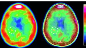
Current visualisation techniques such as magnetic resonance imaging (MRI) can only identify damage to the blood-brain barrier that allow rogue immune cells to enter the brain and trigger self-destructive inflammation, a hallmark of multiple sclerosis. For the first time, Hanna Gerwien and colleagues at the universities of Muenster and Leuven have detected onset of the debilitating and incurable autoimmune disorder in five patients.
The researchers used an imaging probe that binds to matrix metalloproteinases (MMPs), enzymes known to play a central role in MS development, coupled with positron emission tomography (PET) imaging. For the first time, they found in a MS mouse model that MMP activity served as a reliable marker for leukocyte infiltration of the blood-brain barrier – and confirmed their findings in patients.
In all five study participants, the agent penetrated the brain, closely following the infiltrating leukocytes and subsequent inflammation. Lesions detected by this approach correlated with those measured by traditional MRI as well as MMP levels in patients’ cerebrospinal fluid. The results suggest the technology may help doctors spot and diagnose relapsing and progressive forms of MS sooner, as well as observe patients’ responses during anti-inflammatory treatment